How to make radiotherapy more accurate and effective in cancer treatment? Scientists and doctors have joined forces and are working to develop a medical phantom for precise dose planning in radiotherapy. The device will allow both the dose and the angle of incidence of the radiation beam to be tailored to each patient so that healthy tissue is not damaged. The project could lead to a revolutionisation of techniques for measuring radiation dose distribution and personalisation in radiotherapy, which is the most commonly used treatment for cancer patients.
When surgical removal of the tumour is not possible, doctors often opt for radiotherapy. Its aim is to inhibit the further growth of cancer cells and to eliminate them from the body. Radiotherapy is also used as a complementary treatment for patients after surgery. In total, it is estimated that in Poland about half of all patients treated for cancer receive radiation.
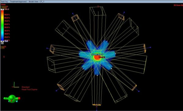
Before the start of treatment with ionising radiation, a precise individual therapeutic plan is created for each patient, specifying exactly the fields to be irradiated, the dose level and the treatment schedule. The planning serves the purpose of hitting the tumour with maximum force while minimising the effect of the rays on healthy tissues, especially on so-called critical organs such as the heart.
In the next step, this plan should be verified. This can be done by independent calculations, measurements or a combination of the two. “Nowadays, with the increasing computing power of computers and the continuous improvement of algorithms for simulating the interaction of ionising radiation with tissues, the precision of the calculation of absorbed dose distributions is getting better, but still the direct measurement of this distribution is considered the best way to verify therapeutic plans,” says Tomasz Szumlak, PhD, professor at the AGH University of Science and Technology in Krakow.
This is why scientists from AGH, the Krakow University of Technology and the Oncology Centre in Krakow formed the Dose-3D consortium and decided to develop an advanced measuring device that will enable a reliable and accurate check of the correctness of the plan in a specific patient. For the research work, the Foundation for Polish Science has provided more than PLN 12 million as part of the TEAM-NET programme.
“Our consortium wants to build a three-dimensional measurement matrix, filled with a tissue-like substance (scintillator), containing a detector capable of directly measuring the spatial distribution of the deposited dose. Such a phantom will be configurable in such a way that it replicates the patient’s body each time,”
– explains Professor Tomasz Szumlak, who leads the consortium’s work.
According to the Dose-3D researchers, their project has the potential to completely change previous methods of measuring radiation dose distribution and creating individualised plans in radiotherapy. In their device, the researchers want to use a liquid, tissue-like scintillator (a substance that emits light when exposed to ionising radiation), instead of the plastic scintillators currently most commonly used in such phantoms.
“The use of active liquid material introduces a number of complications compared to existing solutions, but this will make it relatively easy to relate dose measurements to measurements in patient tissue.”
– said prof. Szumlak
The second innovation will be the use of 3D printing technology to build the detector head. “This approach is still rarely used in the construction of ionising radiation detectors. However, recent developments with 3D printing using carbon seem to be ideal for our project. Carbon is a light element that should not introduce significant secondary effects during detection.” – emphasises the scientist.
Another important component of the project will be the development of high-quality software for dose simulation, configuration and control of the entire device and analysis of the data obtained. Once a prototype of the device has been built, the researchers intend to test it at the Oncology Centre in Krakow and then, assuming this work is successful, to launch pilot production and eventually enter the medical phantom market.
Acknowledgments
The POIR.04.04.00-00-15E5/18 project is carried out within the “TEAM-NET” programme of the Foundation for Polish Science co-financed by the European Union under the European Regional Development Fund.